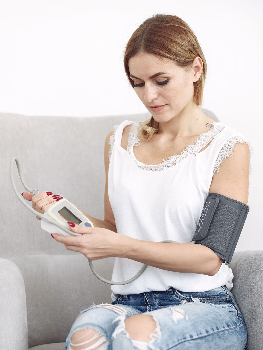
Preeclampsia is a syndrome characterized by high blood pressure, between 3% and 5% of pregnant women can suffer from it and may causes health consequences for both the mother and the developing baby.
There is no clear reason for pre-eclampsia. A part of the hypertensive syndrome of pregnancy, pre-eclampsia is a syndrome that has multiple causes and several mechanisms to produce it. It is classically characterized by elevated blood pressure and the presence of protein in the urine. It appears in the second half of pregnancy (after 20 weeks) but disappears after delivery.
It occurs when the vascular invasion of the trophoblast (cells in the formation stage in the placenta), produces ischemia in the placenta. Endothelium damage and toxins pass into the maternal circulation and affect various organs, such as the kidneys, liver, brain, lungs, pancreas, and vasculature, so the only effective treatment is birth.
It is the main cause of maternal and perinatal morbidity and mortality, the origin of this syndrome is in the placenta, but the primary cause that alters its formation is unknown.
If a pregnant woman has a headache accompanied by visual disturbances, upper abdominal pain or tinnitus (ringing in the ears), progressive and generalized edema, she should immediately consult a doctor, especially if her blood pressure is equal to or greater than 140 / 90 mmHg.
As the placenta is the origin of this syndrome, the only effective and definitive treatment is the interruption of pregnancy. However, this is not always possible, so management is necessary to try to reach fetal maturity and reduce the complications associated with a premature newborn.
DEPENDING ON THE TIME AT WHICH IT IS DIAGNOSED, IT CAN BE CLASSIFIED AS EARLY (BEFORE 34 WEEKS) OR LATE IF PERFORMED AFTER OR ACCORDING TO THE SEVERITY IT PRESENTS
Preventing preeclampsia is difficult. In order to do this in the first trimester, biochemical markers are studied and identified in maternal blood and Doppler measurement of the maternal uterine arteries. Together with the presence of risk factors and alterations in the measurement of blood pressure, have been used for the development and study of early prevention strategies, before 16 weeks, as well as the administration of aspirin, and calcium that will be prescribed during prenatal consultations.
Women who develop pre-eclampsia, especially if it is early or severe, have a higher risk of later developing cardiovascular diseases such as chronic hypertension, strokes, coronary heart disease, diabetes, and kidney failure. For this reason, it is necessary that they continue with controls and make changes in habits to be able to prevent or delay these diseases.
Risk factors for pre-eclampsia :
- First pregnancy
- Age at pregnancy (less than 20 or more than 35 years)
- History of preeclampsia in a previous pregnancy
- Multiple pregnancies
- Obesity
- Family history of preeclampsia (mother and sisters)
- Pre-existing diseases (chronic arterial hypertension, diabetes mellitus, antiphospholipid syndrome, thrombophilia, autoimmune diseases, kidney diseases, infertility)
What does a subchorionic hematoma mean in a pregnancy?
One of the main concerns, in both spontaneous and medically assisted pregnancies, is the presence of bruises or clots near the amniotic sac. They can be divided into recent and old hematomas. As its name indicates, the recent ones are clots that appear as a result of bleeding that occurs in the initial stages of pregnancy. The typical history is a previously healthy patient, who begins to have bleeding, goes to the obstetrician where an ultrasound is performed and the presence of a clot is observed. The findings are determined by two things: first by gestational age (pregnancies less than 12 weeks are more likely to be lost) and the size of the hematoma (bruises greater than 50% in relation to the amniotic sac have a worse prognosis). Treatment is based on absolute rest, assessing the opening of the cervix and the use of medications such as progesterone and indomethacin. However, your GP will indicate which is the most appropriate treatment for your situation.
Old hematomas, on the other hand, are generally incidental findings as the patient usually has no history of bleeding or pain. If a hematoma is found, they are referred to an ultrasound. They are generally due to the rupture of an endometrial vessel that, because it did not cause bleeding through the vagina, went unnoticed. These old bruises can be reabsorbed (that is, disappear on subsequent ultrasound exams) or initiate benign dark bleeding. As in the previous case, the treating doctor will carry out the evaluations and will explain to you what is the best measure for your pregnancy at that time.
Under no circumstances is a hematoma an indication for a curettage. Curettage is performed under two circumstances only: the presence of uncontrollable bleeding that endangers the life of the patient (for example, fainting, changing more than 4 sanitary pads with abundant blood in a couple of hours, etc.) or in the absence of a fetal heartbeat. In early pregnancies, drug management can be performed to expel the bursa (with the embryo already deceased), but each clinical situation and each particular context must be assessed.
We hope this info is useful, and as we have discussed in previous posts, tag whoever needs it and share this information in your stories to create a community supporting women!


