Together with lack of oxygen and prematurity, congenital defects are the main causes of perinatal and infant morbidity and mortality.
In many cases they are the cause of complications and sequelae associated with different degrees of disability that compromise the development and social integration of the individual. A prevalence of 3-6% is estimated. On the other hand, the detection of growth retardation allows a reduction in perinatal mortality, especially that which occurs at the end of pregnancy. These data justify the establishment of protocols aimed at improving prenatal detection rates.
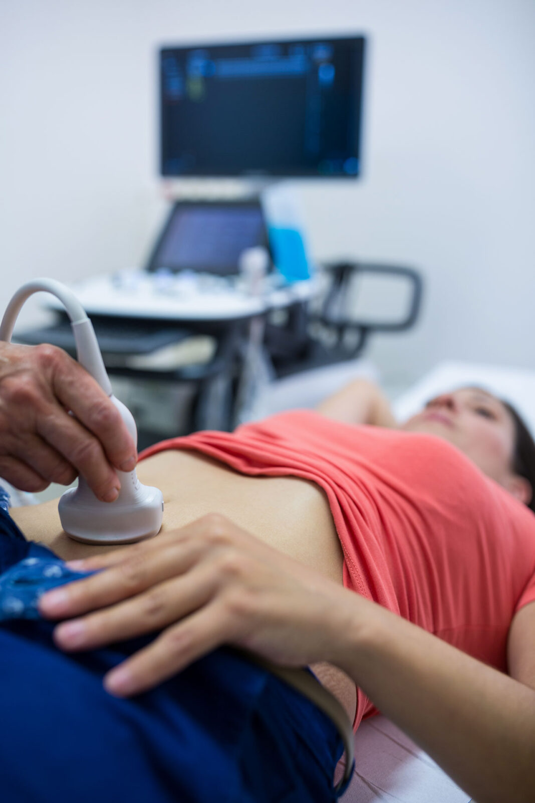
In our UR Maternal-Fetal Department, the care protocol during pregnancy includes performing 3 ultrasounds distributed during the first, second and third trimesters. The timing and objectives of these ultrasounds are summarized below:
I trimester: Week 11 – 13.6. (preferably week 12)
- Confirm intrauterine pregnancy
- Confirm evolution of gestation
- Determine the number of fetuses and chorionicity in case of multiple gestations
- dating the pregnancy
- Determination of aneuploidy markers
- Early anatomical assessment
- Determination of the pulsatility index of the uterine arteries to calculate the risk of preeclampsia.
II Trimester: Week 20 – 22. (preferably week 21)
- Evaluation of placenta, cord insertion and amniotic fluid
- Fetal growth assessment
- Assessment of fetal anatomy
III Trimester: at low risk at 37±1 and at high risk of growth retardation serially at 28±1, 32±1 and 37±1
- Fetal static assessment
- Assessment of placenta and amniotic fluid
- Fetal growth assessment
- Reassessment of the fetal anatomy to rule out evolutionary pathology and/or possible late onset


